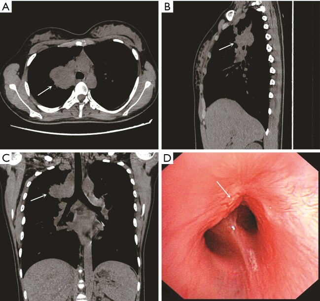
Figure 1.
Computed tomography (CT) of chest and bronchoscopy. (A-C) Transverse/sagittal/coronal planes of chest CT revealed a large mass measuring 4.0×4.8 cm at the proximal of right upper lobe (white arrow); (D) the bronchoscopy revealed that a mass completely obstructed the apical segmental bronchus of the right upper lobe (white arrow).

