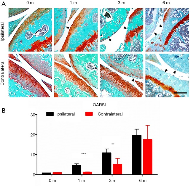
Figure 1.
Ipsilateral and contralateral knees developed OA in distinct patterns in murine ACLT model. (A) Representative photos of Safranin O and fast green staining of ipsilateral (upper) an contralateral side of knee joints 0, 1, 3 and 6 months after ACLT; (B) statistical analysis, n=8 per group, statistical significance was determined by unpaired Student’s t test and all data were shown as a bar with means ± standard deviations. Scale bars, 500 µm. **, P<0.01, ***, P<0.001 compared with the sham operated group at different time points. ACLT, anterior cruciate ligament transection; OA, osteoarthritis.

