
Figure 1.
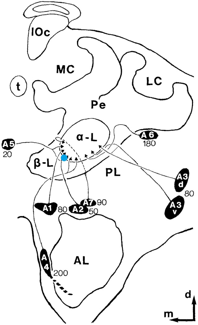
Recording site. Schematic drawing of one hemisphere of the honeybee brain. The blue dot depicts the recording site. MB ENs (mushroom body extrinsic neurons)of the alpha-lobe A1, A2, and A5 were targeted. The black ovals with the names of the neuron clusters mark the respective somata regions. The depth of theses neurons is indicated by the numbers near the clusters. The outer edge of the alpha-lobe can be recognized very well as a guide for electrode placement (adopted from Rybak and Menzel, 1993; Figure 3).