
Figure 2.
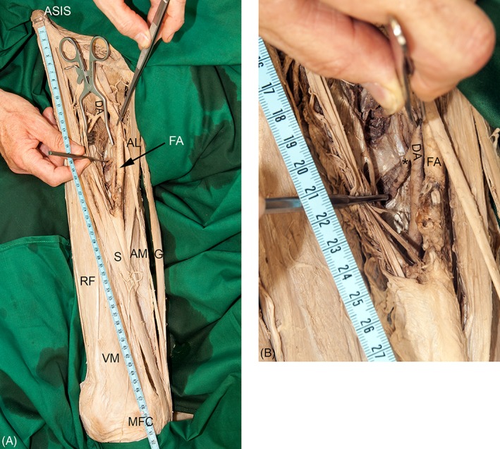
A‐B The photographs exemplify the measurement of the passage of one perforating artery (asterisk). (A) The image shows the reference line between the anterior superior iliac spine (ASIS) and the medial femoral condyle (MFC). (B) The photograph gives a detailed view of the measurement using a probe. AL, adductor longus muscle; AM, adductor magnus muscle; DA, deep artery of thigh; FA, femoral artery; G, gracilis muscle; RF, rectus femoris muscle; S, sartorius muscle; VM, vastus medialis muscle. [Color figure can be viewed at http://wileyonlinelibrary.com]