
Figure 6.
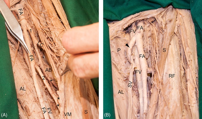
(A) The picture shows an example with two deep arteries of thigh instead of one in the left leg of a female individual. The regular deep artery of thigh (1) gave rise to the first perforating artery (asterisk). From the aberrant deep artery of thigh (2), the remaining perforating arteries (asterisks) arose. (B) In the left leg of a female individual, the deep artery of thigh (arrowhead) looped superficially around the femoral vein (FV) and great saphenous vein (SV). This situation may lead to problems during puncture or endovascular procedures of the femoral artery (FA) or FV. AB, adductor brevis muscle; AL, adductor longus muscle; P, pectineus muscle; RF, rectus femoris muscle, VM, vastus medialis muscle; S, sartorius muscle. [Color figure can be viewed at http://wileyonlinelibrary.com]