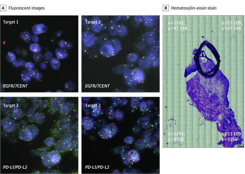
Figure 3. Patient 37.
This patient had a pT2N3a Lauren intestinal subtype adenocarcinoma arising from the gastric cardia. OncoScan analysis revealed major copy number alterations of CD274, PDCD1LG2, EGFR, MET, and PIK3CA. A, Target 1 image at coordinates x = 13 654 μm and y = 33 594 μm exhibited modest copy number gains of EGFR. Target 2 images at coordinates x = 11 292 μm and y = 34 466 μm exhibited amplification in all 5 oncogenes. B, The tumor area of interest is circled on the hematoxylin-eosin–stained (H&E) slide section with the x, y reference coordinates at the 4 corners of the image displayed in micrometer distances. All fluorescent images were obtained at magnification x60, and whole H&E tumor slide section image, magnification x5.

