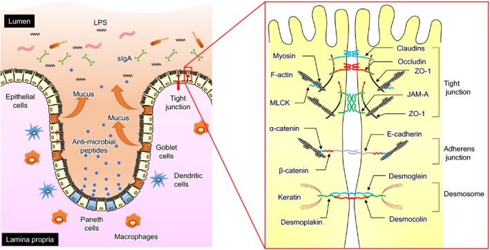
FIGURE 1.

Anatomy of the intestinal barrier. Intestinal epithelial cells constitute a biochemical and physical barrier to the diffusion of pathogens, toxins, and allergens from the intestinal lumen to the mucosal tissues (left panel). The intestinal barrier system depends on interactions among several barrier components, including the adhesive mucous gel layer, immunoglobulin A, antimicrobial peptides, and intercellular tight junctions (TJs). TJs are formed by a multiple‐protein complex located in the apical portion of the lateral membrane of epithelial cells (right panel). The TJ structure comprises transmembrane proteins, such as claudin, occludin, and junctional adhesion molecule‐A (JAM‐A), and intracellular plaque proteins, such as zonula occludens (ZOs) and cingulin. The interaction between extracellular regions of the transmembrane proteins in adjacent cells regulates the paracellular passage of molecules
