Figure 1.

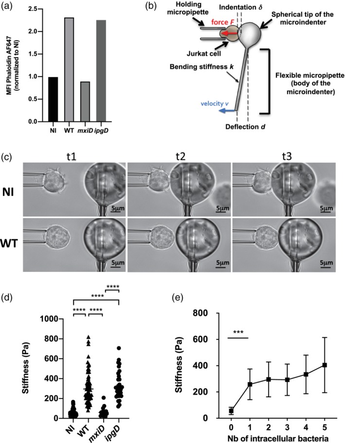
Increased stiffness associated with increased filamentous actin in Shigella‐invaded Jurkat cells. (a) F‐actin content measurement. Cells were either non‐infected (NI) or infected with WT‐Shigella or the mxiD or ipgD mutants. Cells were fixed and permeabilised prior to staining with fluorescent phalloidin whose intensity was analysed by flow cytometry. Representative of three independent experiments. (b) Principle of the profile microindentation setup. A stiff micropipette holds a cell. A flexible micropipette has a glass sphere at its tip and is used as a microindenter, with its base translated towards the cell at a constant velocity (v). Upon contact with the cell, the glass bead induces a deformation of the cell (refers as the indentation δ) allowing calculation of the cell stiffness. (c) Representative snapshots to illustrate the microindentation experiment at three randomly chosen time points (referred as t1, t2, and t3). Representative NI cell (upper panel) and wild‐type (WT) Shigella‐invaded cell (bottom panel) are represented. (d) Stiffness measurements in NI, mxiD‐infected cells, WT‐ or ipgD‐invaded cells (three independent experiments, except for the mxiD strain, two independent experiments). (e) Impact of the number of intracellular WT Shigella on cell stiffness from six independent experiments. (a,d,e) M ± SD, ***p < .001, ****p < .0001 (Kruskal–Wallis and Tukey's multiple comparisons test)
