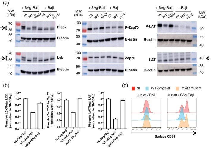
Figure 4.

Shigella prevents human Jurkat T cells activation. Jurkat cells were infected with wild‐type (WT) Shigella or the mxiD mutant for 30 min at a multiplicity of infection of 20 followed by 1 h30 gentamicin to kill extracellular bacteria. (a) Following sorting of Shigella‐invaded Jurkat cells, these cells were then incubated with Raji or SAg‐Raji cells for 20 min and lysed. Supernatants were analysed by immunoblotting using individually specific antibodies against Lck, ZAP70, and LAT and their phosphorylated forms. β‐Actin immunoblotting used as a loading control for each blot is shown. The arrows indicate the bands used for quantification in Figure 4b. (b) Ratio of phosphorylated protein over total for Zap70, Lck, and LAT when Jurkat cells were incubated with SAg‐Raji cells. One representative experiment is shown. (c) Jurkat cells were stimulated with Raji cells pulsed (SAg‐Raji) or not for 2h30 min. Cells were fixed and stained with anti‐CD69 mAb. Surface expression of CD69 on live cells from one representative experiment out of three is shown. NI, non‐infected
