Figure 2.

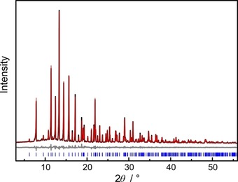
Rietveld refinement of BaP6N10NH; observed (black) and simulated (red) powder X‐ray diffraction patterns and difference profile (grey). Positions of Bragg reflections of BaP6N10NH (blue) are marked with vertical blue bars.

Rietveld refinement of BaP6N10NH; observed (black) and simulated (red) powder X‐ray diffraction patterns and difference profile (grey). Positions of Bragg reflections of BaP6N10NH (blue) are marked with vertical blue bars.