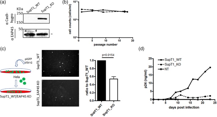
Figure 2.

Depletion of EAP45 impairs virus spreading in SupT1 cells. (a): Western blot of the cell lysates from SupT1 WT and SupT1 EAP45 KO cells. Asterisks show the band of interest, whereas the “ ∼” indicates nonspecific products from the antibody binding on the membrane throughout this study. The size markers are shown to the left of the blot. (b): The number of SupT1 and SupT1 EAP45 KO cells was counted during 18 passages and was plotted against passage number. (c): HIV GFP virus peudotyped with VSV‐G was produced in Hela cells before transduction into either SupT1 WT or SupT1 EAP45 KO cells. The number of GFP positive cells produced from the SupT1 EAP45 KO cells was normalised against those from the SupT1 WT cells. Error bars represent the standard error of the mean from three independent experiments with a statistical significance shown by one sample t‐test (p = .0154). (d): Two million of either SupT1 WT or SupT1 EAP45 KO cells were infected with replication competent HIV BH10. The supernatants were collected every 2–3 days and quantified by p24 enzyme‐linked immunosorbent assay. The plot is representative of two experiments
