
Figure 2.
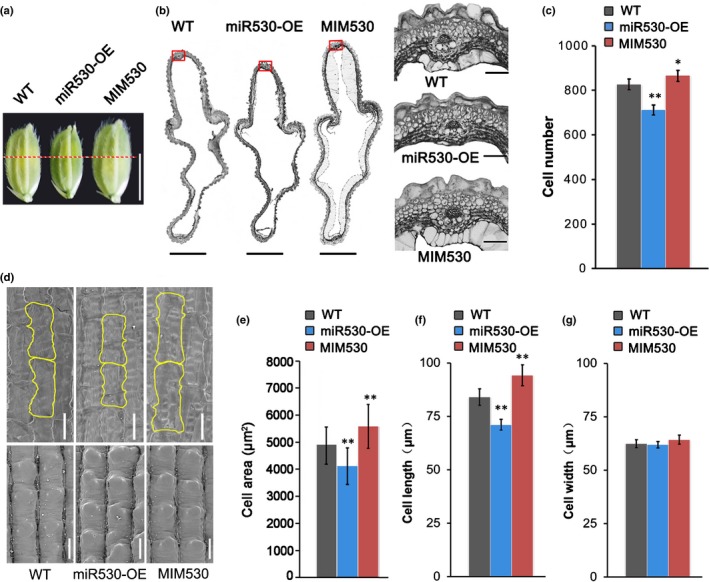
Histological analysis of spikelet hulls in OsmiR530 transgenic lines and wild‐type (WT) plants (miRNA, microRNA; Os, Oryza sativa). (a) Spikelet hulls of rice WT and transgenic plants just before anthesis. Bar, 5 mm. (b) Cross‐sections of the middle parts of spikelet hulls (marked by red dashed lines in a) of rice WT and transgenic plants just before anthesis (left). Bars, 500 μm. Magnified images of the boxed cross‐section areas are provided on the right. Bars, 50 μm. (c) Number of cells in the outer parenchyma layer of the spikelet hulls of rice WT and transgenic plants (n = 5 spikelets). (d) Scanning electron micrographs of the inner epidermal cells of lemmas in the mature seeds of rice WT and transgenic plants. Bars, 50 μm. (e) Cell areas in the middle part of the inner epidermis of lemmas in fully mature rice seeds (n = 5 seeds). (f) Cell lengths in the middle part of the inner epidermis of lemmas in fully mature rice seeds (n = 5 seeds). (g) Cell widths in the middle part of the inner epidermis of lemmas in fully mature rice seeds (n = 5 seeds). The data in (c), (e), (f) and (g) are presented as the mean ± SD. Significant differences: *, P < 0.05; **, P < 0.01 (Student's t‐test).