Figure 1.

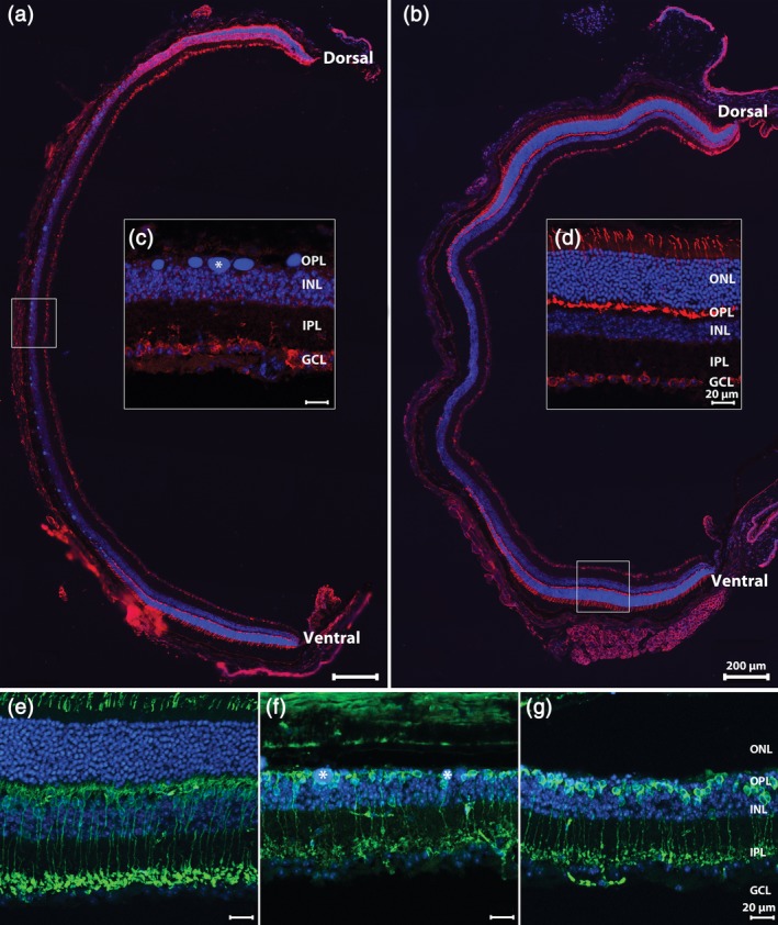
Dorsoventral, longitudinal sections of Tvrm4 retinas 3 (a) and 6 (b) weeks after photoinduction, stained for anti‐Cone Arrestin (red) and Hoechst nuclear marker (blue). (c) During this time interval, photoreceptor (PR) degeneration becomes nearly complete in the central retina where DNA clusters (asterisk) are leftovers from PR nuclear condensation. At both time points examined, intact PRs can be found in more retinal regions, adjacent to the area where the ONL is totally missing, as highlighted in (d). (In this and other pictures: GCL, ganglion cell layer; INL, inner nuclear layer; IPL, inner plexiform layer; ONL, outer nuclear layer; OPL, outer plexiform layer.). Vertical sections of the Tvrm4 retina without photoinduction (e) or 3 (f) and 6 (g) weeks postinduction (PI) stained for anti‐PKCα (green) and Hoechst nuclear marker (blue). In the areas devoid of photoreceptors (f,g) rod bipolar cells retract dendrites and their axonal endings exhibit a visible shrinkage (g) [Color figure can be viewed at http://wileyonlinelibrary.com]
