
Figure 4.
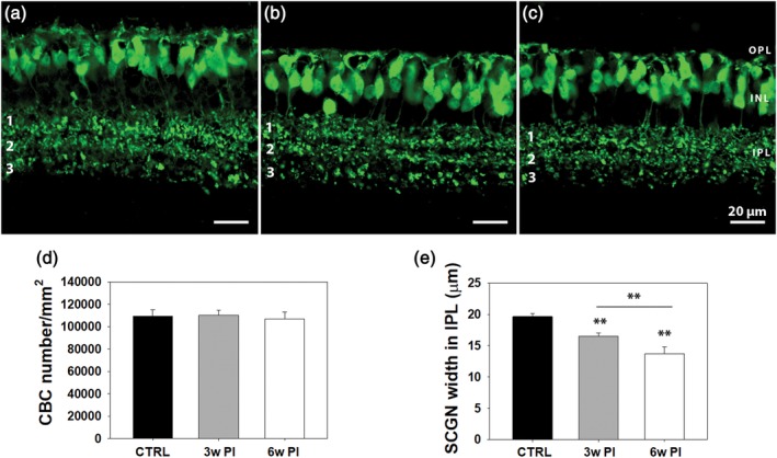
Vertical sections from retinas of WT controls (a) and Tvrm4 mice, 3 (b) and 6 (c) weeks postinduction (PI), with secretagogin (SCGN) staining of cone bipolar cells (CBCs). Three distinct plexa (1, 2, 3) of CBC axonal arbors are visible in the IPL. A gradual, significant decrease in the thickness of SCGN+ axonal arborizations becomes evident over time, mostly due to plexus 3 shrinkage, despite preservation of total IPL width. (d,e) CBC quantitative data. Three vertical sections/animal were analyzed taking 10 micrographs/section, covering five fields, each 150 × 150 μm2. Cell body counting reveals no statistical difference in control and Tvrm4 cases (d). IPL thickness measurements were done on the same micrographs with a MetaMorph longitudinal measuring tool (10 random measurements/micrograph done along the full IPL thickness in the vertical plane (** = p ≤ .001; data are shown as average and SE; one‐way analysis of variance [ANOVA]) [Color figure can be viewed at http://wileyonlinelibrary.com]