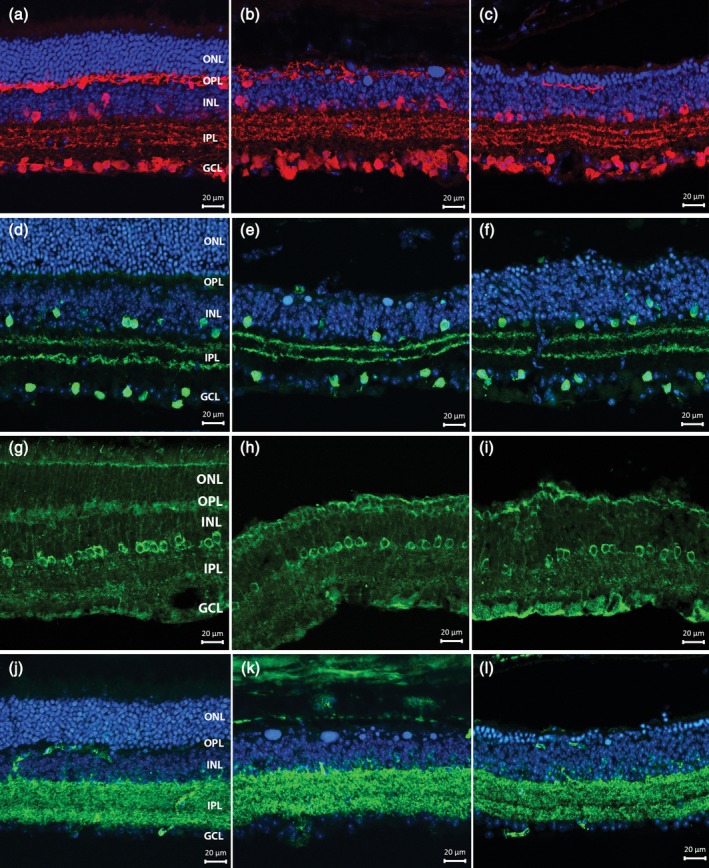
Figure 5.

Vertical sections from noninduced control (left column) (a,d,g,j), 3 weeks (central column) (b,e,h,k) and 6 weeks (third column) (c,f,i,l) Tvrm4 retinas. Antibody stainings highlight amacrine cell morphologies and organization: (a–c) Calbindin (red); (d–f) ChAT (green); (g–i) DAB1; and (j–l) GAD67. Blue: Hoechst nuclear counterstaining. The pattern of immunoreactivity in the inner retina is similar to the control at both time points. No major changes are observed in the general morphology and laminar organization of stained bodies and processes [Color figure can be viewed at http://wileyonlinelibrary.com]
