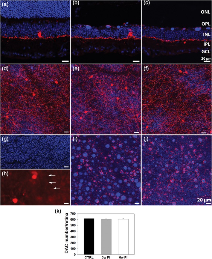
Figure 6.

Vertical retinal sections of Tvrm4 mice stained with anti‐TH (red) and Hoechst (blue) in a non‐induced control eye (left column) (a) and 3 weeks (b) or 6 weeks (c) following light exposure. (d–j) Whole mount micrographs of Tvrm4 retinas as above. The focal plane of (d–f) is in the IPL; (g–j) are taken at the ONL/OPL border. (g,h) The inner limit of the ONL and the outer limit of the OPL, respectively, with arrows pointing at TH‐positive processes in the outer retina of a control mouse. In induced retinas (e,f,i,j), the inner plexus (e,f) originating from DACs shows a normal morphology while processes in the OPL are hypertrophic and denser (i,j). Exuberant processes are observed entangling DNA bodies residual from dead PRs. (k) Counts of DAC+ cell bodies (3 retinas/exp. group; counts performed on tiled images of retinal whole mounts) revealed no significant changes 3 and 6 weeks PI. Tvrm4 data are compared to the published number of DACs in WT B6/J (CTRL) mice (Whitney, Raven, Ciobanu, Williams, & Reese, 2009) (data are shown as average and SE; one‐way analysis of variance [ANOVA]) [Color figure can be viewed at http://wileyonlinelibrary.com]
