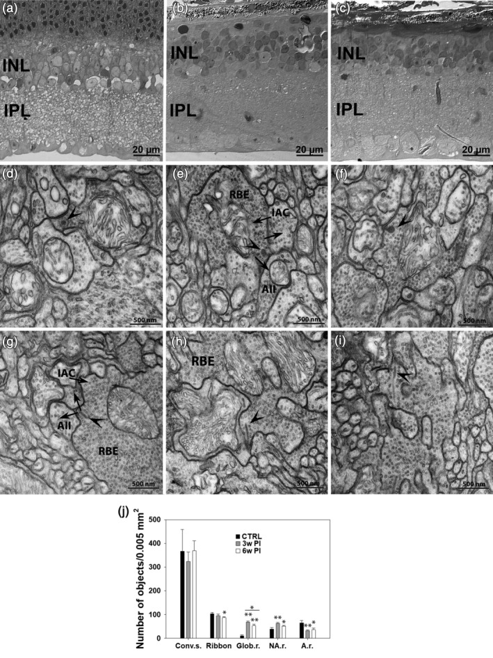
Figure 8.

Semithin vertical sections of Translational Vision Research Model 4 (Tvrm4) mouse retina stained with toluidine blue in a noninduced, control eye (a) and 3 (b) and 6 weeks (c) following light exposure. (d–i) TEM micrographs illustrating synaptic ribbons (arrowheads) and conventional chemical synapses (arrows) involving rod bipolar axonal endings (RBE) in a noninduced control retina (d,g) and in Tvrm4 preparations examined 3 (e,h) and 6 (f,i) weeks postinduction (PI). Arrows indicate the direction of neurotransmission. Atypical globular ribbons (d–f, arrowheads) and classical synaptic arrangement involving RBE with elongated ribbons and dendrites of AII and A17‐like amacrine cells (IAC) can be observed in all preparations (e–i). A summary of conventional and ribbon synapse density in the IPL is shown in (j). TEM micrographs (400/sample, 12.96 μm2/micrograph)/sample, corresponding to a sampled area of 0.0052 mm2/retina) were imaged). The number of conventional synapses (Conv. s.) is not affected by photoinduction. The total number of ribbons shows a significant decrement 6 weeks PI (* = p ≤ .01; one‐way analysis of variance [ANOVA]). Also, significantly more ribbons have a globular shape (Glob. r.) (** = p ≤ .001; one‐way ANOVA), 3 and 6 weeks PI, compared to non‐induced controls (CTRL). Globular ribbons represent 72% of all ribbons 3 weeks PI but decrease to 61% 6 weeks PI. The number of nonanchored ribbons (NA. r.) is significantly higher 3 and 6 weeks PI, while the number of anchored ribbons (A.r.) shows the opposite tendency. All data are shown as average and SE
