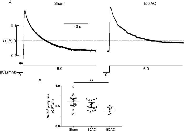
Figure 7.

The function of the Na+, K+ ATPase, following pump reactivation, between experimental groups
A, when the Na+,K+‐ATPase was inhibited by superfusion of the cells in K+‐free solution, the [Na+]i is expected to increase. After 2 min the superfusate was switched to NT containing 6 mm K+ to reactivate the Na+,K+‐ATPase. Reactivation extrudes the accumulated Na+ from the cell and this rate of extrusion was used to assess the function of the Na+,K+‐ATPase. B, pump current measured in myocytes isolated from hearts at 150 days post‐AC (150AC) had reduced Na+ extrusion rates compared with the sham group. There was no change in Na+ extrusion rates in the age‐matched sham myocytes so these data were combined. One‐way ANOVA with a Sidak post hoc test ** P < 0.01; n = 18, 15 and 7 for sham, 60AC and 150AC, respectively from 4–8 hearts. Scatter plots show mean (95% CI).
