
Figure 13.
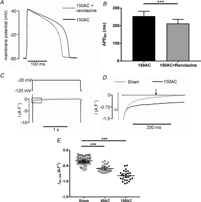
Changes in INa, late during progression towards HF
A, APD was shortened in the presence of 10 μm ranolazine in a cardiac myocyte from the 150AC group of animals. B, mean data showing APD90 in cardiac myocytes from 150AC group was significantly shortened in the presence of 10 μm ranolazine (150AC, n/N = 23/6; mean (SD), Student's t test, ** P < 0.001). C and D, typical I Na,late traces in 150Sham and 150AC cells. E, scatter plot of pooled data with mean (95% CI) for each group. There were no changes in I Na,late in the age‐matched sham myocytes so these sham data were combined. *** P < 0.001; n = 58, 27 and 26 from 4–8 hearts for Sham, 60AC and 150AC respectively. One‐way ANOVA with a Sidak post hoc test.