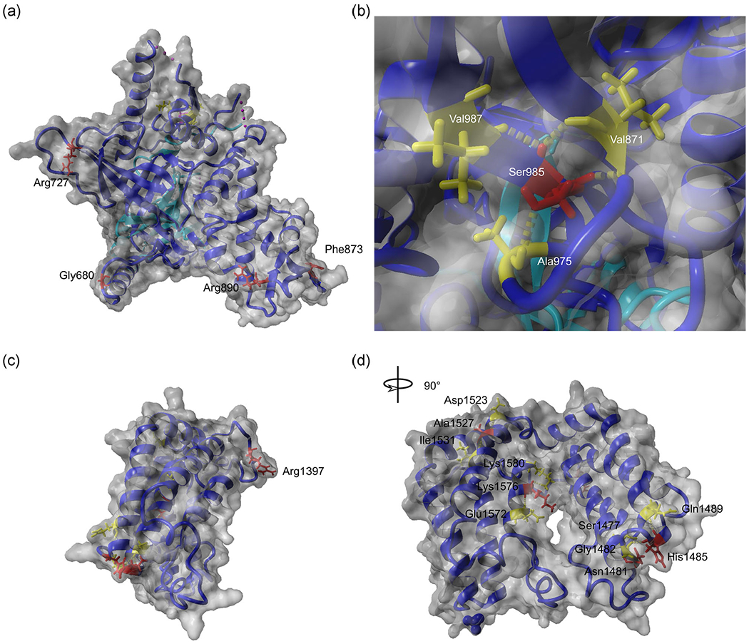
FIGURE 3.

Molecular modeling of TAF1 variants. TAF1 mutations located in the TAF1-TAF7 interaction domain are mapped in the crystal structure deposited by Wang et al. (2014) (PDB: 4RGW) (a,b). Mutations located in the double BrDs are mapped using the structure deposited by Jacobson et al. (2000) (PDB: 1EQF) (c,d). The TAF1 molecule is depicted in blue, the TAF7 molecule in cyan (a,b). TAF1 residues altered in patients with intellectual disability are indicated in red and their interacting amino acids are in yellow. BrD, bromodomain; TAF1, TATA-box binding protein associated factor 1
