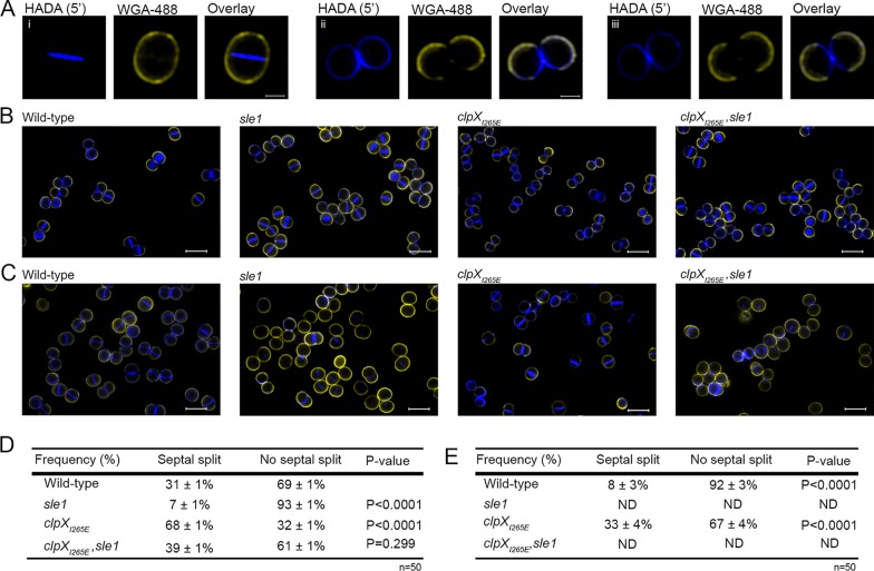
FIG 3.
High Sle1 levels accelerate splitting of S. aureus daughter cells, whereas inactivation of Sle1 and oxacillin delay splitting of fully divided daughter cells. SR-SIM images of exponential cells of the JE2 wild-type, JE2sle1, JE2clpXI265E, and JE2clpXI265E,sle1 strains grown in the absence (A and B) or presence of oxacillin (C) (0.05 μg ml−1 for 20 min). Prior to imaging, cells were stained for 5 min with WGA-488 (green) and HADA (blue). (A) Sample images of cells that have completed septum synthesis during the labeling period (closed septal HADA signal) and that have either not separated after labeling (i) or have initiated daughter cell separation after labeling (ii and iii). (D and E) Septal splitting was quantified in 50 cells displaying a closed HADA-stained septum in cells not exposed to oxacillin (D) and in cells exposed to oxacillin (E). The experiment was performed in two biological replicates, and a statistical analysis was performed using a chi-square test comparing either the splitting frequency between mutant and wild-type cells (D) or between cells not exposed or exposed to oxacillin (E).

