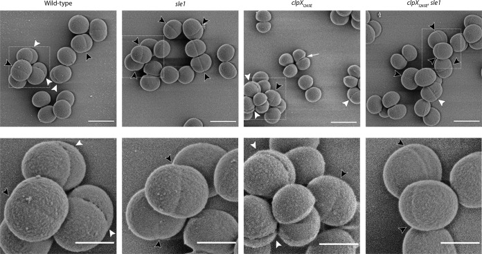
FIG 5.
Perforations in the peripheral septal wall correlate with Sle1 levels. SEM images of JE2 wild-type, JE2clpXI265E, JE2sle1, and JE2clpXI265E,sle1 strains grown in TSB to mid-exponential phase at 37°C. Black arrowheads indicate cells displaying invaginations without cracks at midcell; white arrowheads point to cells displaying cracking along the septal ring at midcell. The white arrow points to a JE2clpXI265E cell that has initiated daughter cell splitting. Boxes in the upper panel are enlarged in the lower panel. Scale bars, 1 μm (upper panel) and 0.5 μm (lower panel).

