
Figure 3.
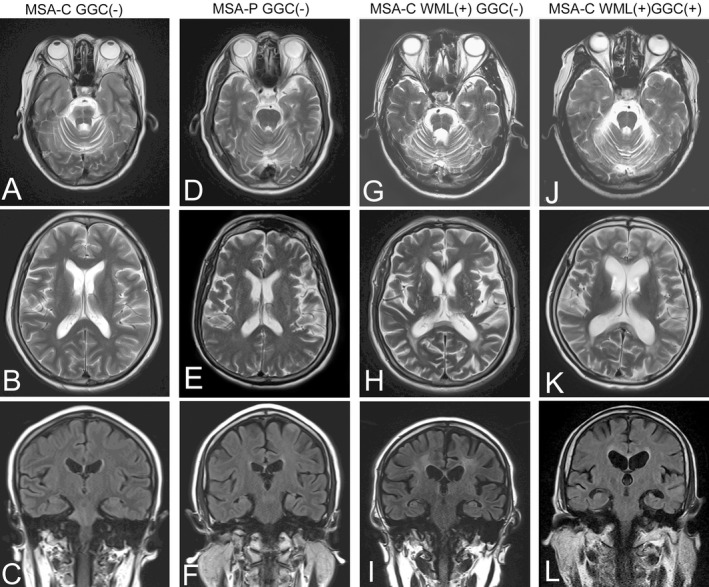
Magnetic resonance imaging representative in this group of MSA patients. Cerebellar atrophy and hot cross bun sign were indicated in a MSA‐C patient without GGC expansion (A–C). Putamen atrophy and lineal T2 high intensity of the lateral margin of the putamen were observed in a MSA‐P patient without GGC expansion (D–F). White matter lesions (WML) suggesting chronic ischemic arteriopathy were notable in a MSA‐C patient without GGC expansion (G–I). White matter lesions and cortical atrophy were found in a MSA‐C patient with GGC expansion (J–L).