Figure 1.

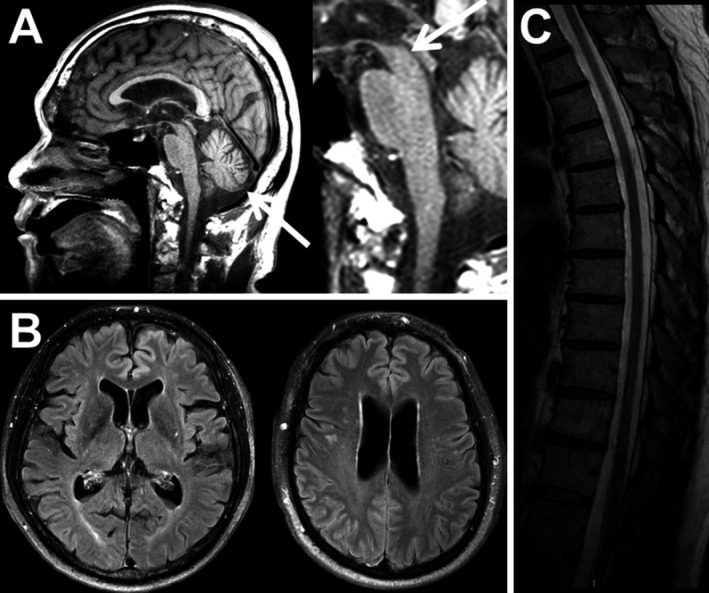
CNS neurodegeneration in Erdheim–Chester Disease. (A) Cerebellar and midbrain atrophy seen on FLAIR imaging. (B) Cerebral atrophy (FLAIR imaging). (C) Spinal cord atrophy (T2‐weighted image).

CNS neurodegeneration in Erdheim–Chester Disease. (A) Cerebellar and midbrain atrophy seen on FLAIR imaging. (B) Cerebral atrophy (FLAIR imaging). (C) Spinal cord atrophy (T2‐weighted image).