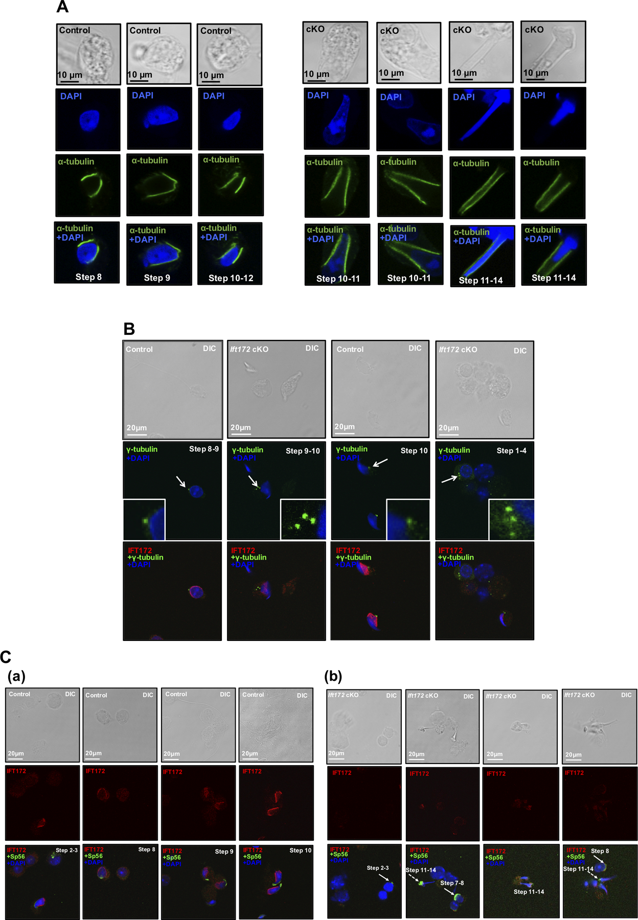
Figure 6. Knockout of Ift172 leads to abnormal manchette morphology and centrosomes but normal acrosome biogenesis in germ cells.

Isolated germ cells from control mice and conditional Ift172 KO mice were stained with indicated antibodies.
(A) IF was conducted with an anti-α-tubulin antibody that labeled manchette of elongating spermatids (in green). Nuclei were stained in blue (DAPI). Notice that the manchette of cKO mice (right panel) appeared to be longer than that in the control mice (left panel);
(B) IF was conducted with the IFT172 antibody (in red) and a centrosome marker, γ-tubulin (in green). Notice that multiple centrosome signals were discovered in the Ift172 cKO male germ cells (white arrows).
(C) IF was conducted on germ cells isolated from the control (a) and Ift172 cKO mice (b) with the anti-IFT172 antibody (in red) and an acrosome marker, Sp56 (in green). Notice that Sp56 localization was normal in round spermatids (white arrows), but appeared to be abnormal in some elongating spermatids in the Ift172 cKO mice (white dashed arrows).
