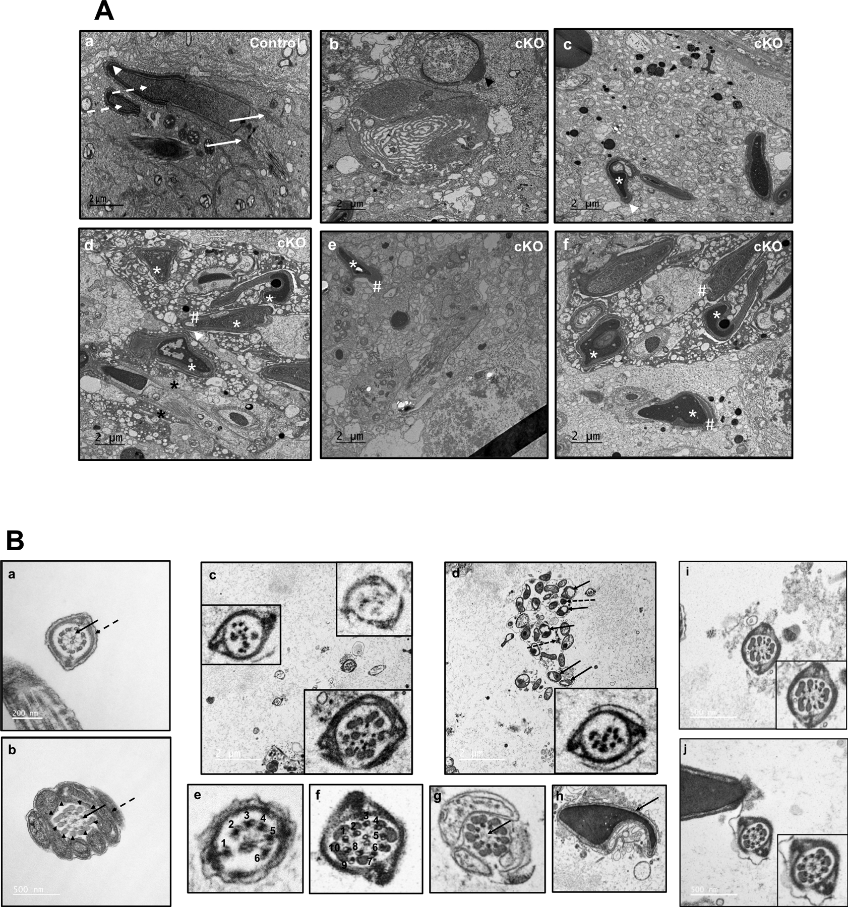
Figure 7. Analysis of testis and epididymal sperm of the control and Ift172 cKO mice by transmission electron microscopy (TEM).

(A) Representative TEM images of testes from control (a) and conditional Ift172 KO mice (b to f). Normally formed manchette (arrows), heads (dashed arrows) and acrosome (arrow heads) were present in the control mice. However, in the cKO mice, a number of abnormalities were observed, including abnormal head formation (white * in c-f), mislocalized and longer manchette (black *). The acrosome was normal in the round spermatid (b), but distorted in elongating spermatids, probably secondary to the abnormal heads (# in d-f).
(B) Representative TEM images of epididymal sperm. Sperm from the control mice (a, b) have normal “9+2” axonemal microtubule cores (arrows) and accessary structures (dashed arrows and arrow heads). (c-j): TEM of sperm from the Ift172 cKO demonstrate multiple abnormalities in both the core axonemes and accessory structures. (c) The left and right lower inserts show two flagella with disrupted core structures; the right upper insert shows a flagellum with almost no internal structure. (d) The arrows point to mitochondria, and the dashed arrows point to disrupted unit fibers of the outer dense fiber (ODF) that are not assembled into the flagellum. The insert illustrates another flagellum with disrupted internal structure. (e) This image represents a flagellum with only 6 outer microtubules. (f) This photo shows a flagellum with ten outer microtubules. (g) In this cross section of the midpiece, the flagellum has normal mitochondria and ODF, as well as nine outer doublet microtubules in the axonemal complex, but the central apparatus (arrow) is missing the two microtubules and associated linking proteins. (h) This is the head of a spermatozoon (arrow) with an abnormal nuclear shape. (i) The flagellum shows a normal fibrous sheath, but a disorganized axonemal core structure. (j) The flagellum shows a normal fibrous sheath and core axoneme structure, but there is a reduction in the ODF subunits.
