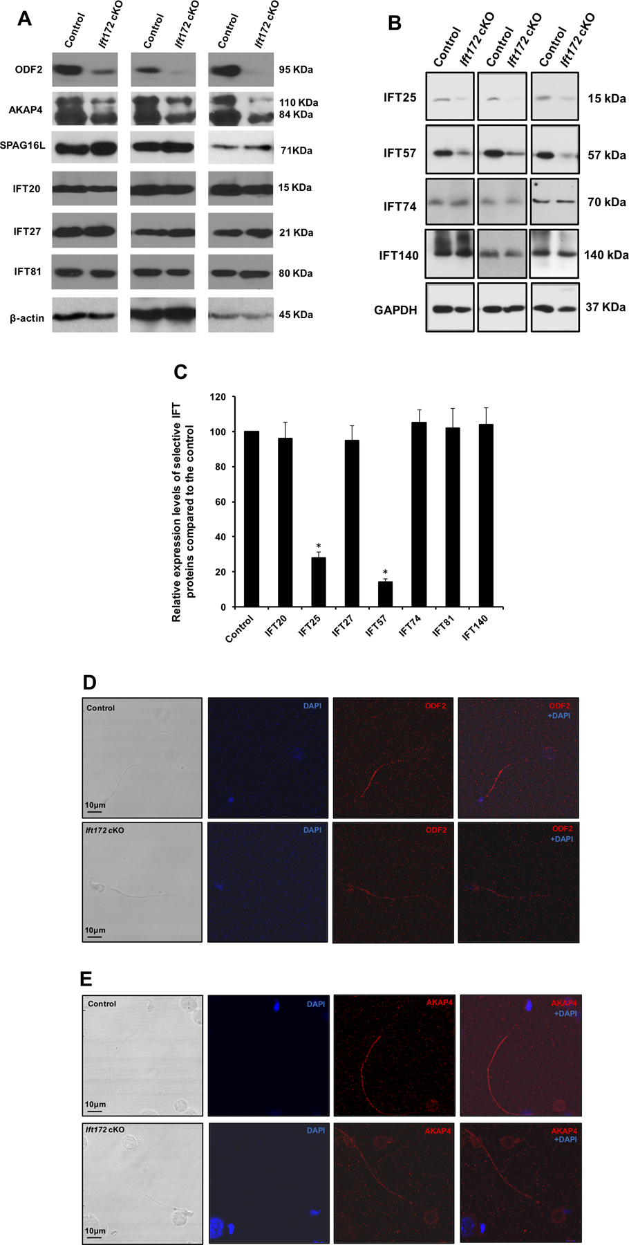
Figure 8. Expression of ODF2, AKAP4, SPAG16L and selective IFT proteins in the conditional Ift172 KO mice.

(A) Representative Western blotting images showing testicular expression levels of three major components of sperm tail (ODF2, AKAP4 and SPAG16L), and three subunits of the IFT system (IFT20, IFT27 and IFT81) using specific antibodies in three control and three conditional Ift172 KO mice. Disruption of Ift172 expression decreased the level of ODF2 and AKAP4, while it had little effect on the expression levels of SPAG16L and three IFT proteins. β-actin was used as an internal control.
(B) Representative Western blotting images of another four IFT proteins. Notice that expression levels of IFT25 and IFT57 in the cKO mice were lower than that in the control mice.
(C) Statistical analysis of relative expression of the IFT proteins normalized by β-actin or GAPDH. There is no difference in expression levels of IFT20, IFT27, IFT74, IFT81 and IFT140 between the control and the Ift172 cKO mice. IFT25 and IFT57 expression levels are significantly reduced in the Ift172 cKO mice.
(D) & (E) Representative images of localization of ODF2 and AKAP4 in the epididymis sperm of a control mouse and an Ift172 cKO mouse, respectively. Both ODF2 and AKAP4 signals were reduced in the Ift172-null sperm flagella.
