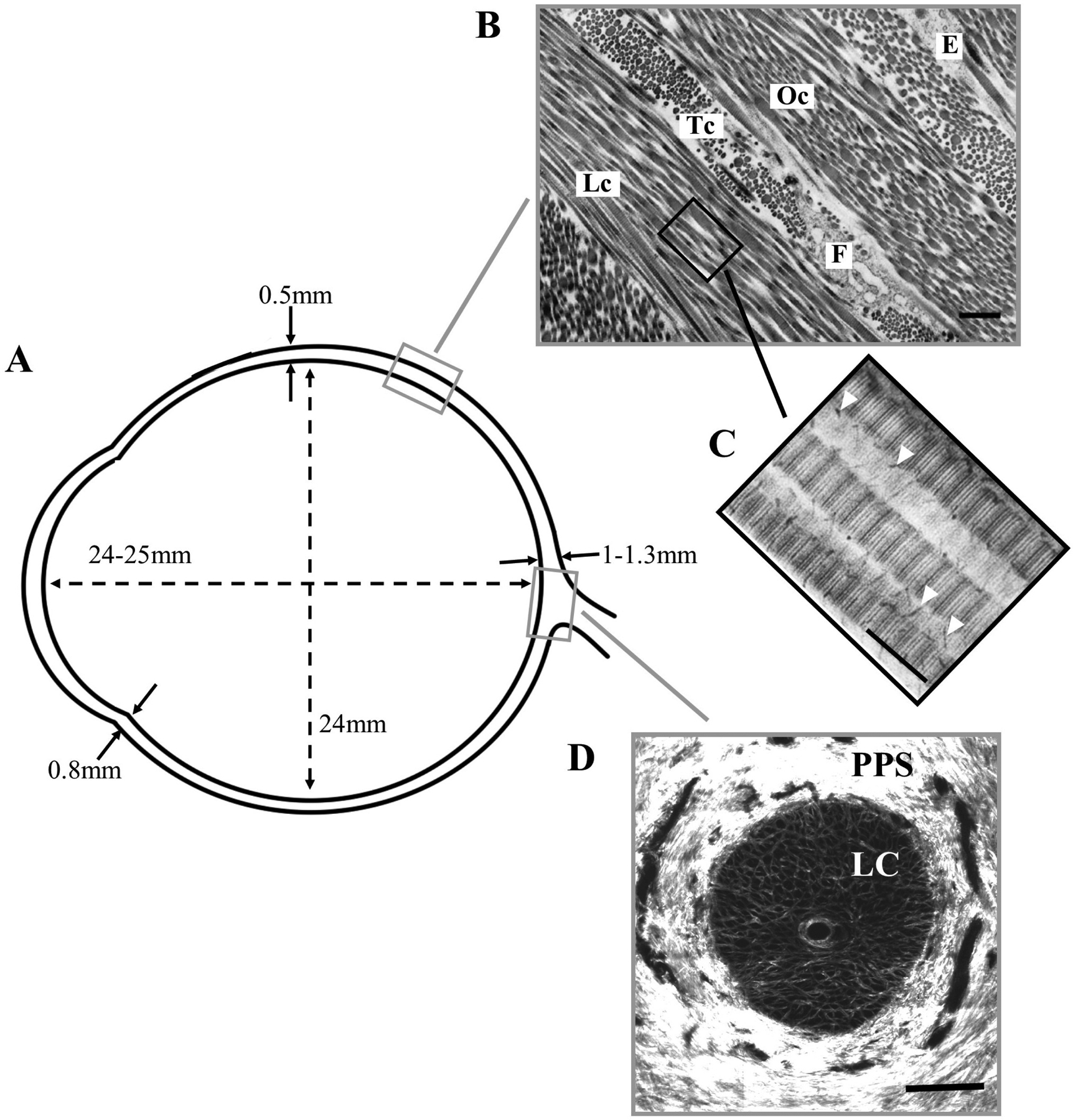
Figure 1:

Overview of scleral morphometry and connective tissue structure. A) Approximate wall thickness and dimensions of the normal, adult human sclera. B) Transmission electron microscopy (TEM) image of the outer scleral stroma, showing lamellar structure formed by collagen fibril bundles in longitudinal (Lc), transverse (Tc) and oblique (Oc) section. A fibrocyte (F) and elastin fibre (E) can also be seen. Bar: 1.5μm. C) TEM image of stroma from a different specimen at higher magnification, showing D-periodic banding of individual fibrils in longitudinal section. Proteoglycans are present as fine filaments (arrowheads) associated with the collagen fibrils. Bar: 250nm. D) Second harmonic generation (SHG) image of en-face section through the optic nerve head at mid-stromal depth, showing the fenestrated lamina cribrosa (LC) that supports the exiting retinal nerve axons. The collagen fibril bundles of the neighbouring peripapillary sclera (PPS) adopt a predominantly circumferential orientation in this region. Panel B taken from (Bron et al., 1997) and reproduced with permission of Hodder Arnold. Panel C adapted from (Watson and Young, 2004) with permission of Elsevier Ltd.
