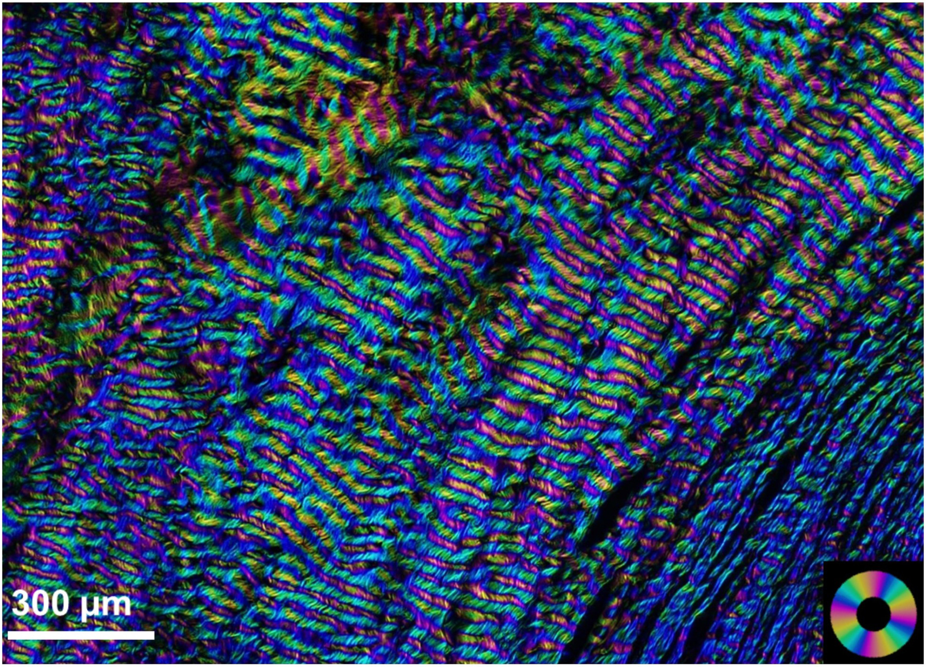
Figure 16:

Porcine PPS imaged by snapshot polarized light microscopy (Yang et al., 2019). The colors indicate the local orientation of the collagen fibres and the brightness is roughly proportional to the local collagen density. Note that the colors are obtained through optical means, and the image is not coloured digitally. The scleral canal is slightly out of frame on the bottom right corner. Clearly discernible in the image are collagen fibre bundles circumferential to the canal. The width of the region of circumferential fibres is between 20% and 40% of the canal diameter in both porcine and human eyes (Gogola et al., 2018b). It is also possible to distinguish the collagen fibres that form the bundles. The bands of color indicate collagen fibre crimp (Jan et al., 2017b). The collagen fibre bundles and the crimp of the fibres increase in size with distance from the canal (Jan et al., 2017a).
