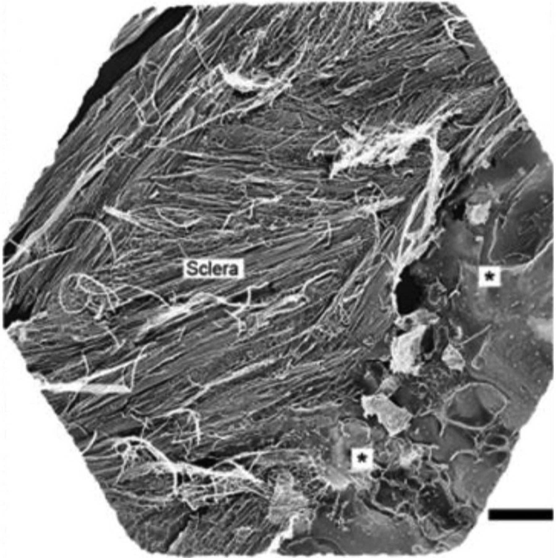
Figure 18:

Quick-freeze deep-etch (QFDE) electron microscopy image of mouse posterior sclera, revealing layers of differentially oriented collagen lamellae in 3D. On the right side of the image (*) can be seen an area of partially etched, vitrified ice - a product of the “freeze-fracture” processing that can preserve native hydrated structure more closely than is possible with conventional electron microscopy sample preparation. Scale bar: 2 μm. Figure reproduced from (Ismail et al., 2017) with permission of Elsevier Ltd.
