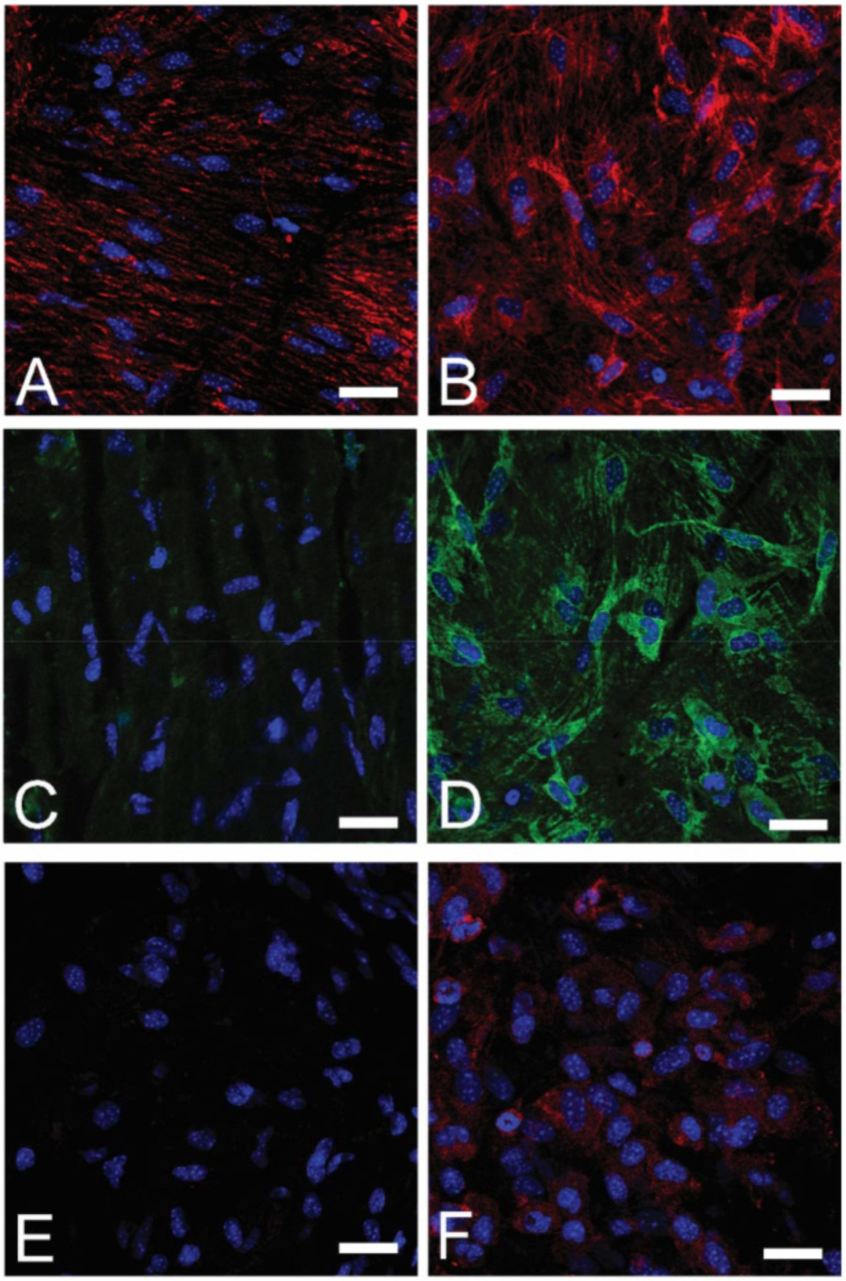
Figure 23:

Experimental glaucoma increases cell proliferation and myofibroblast differentiation in mouse sclera. Top row: Immunohistochemical labelling of vimentin (red) in A) control and B) 3-day glaucoma scleral wholemounts. Middle row:→SMA labelling (green) in C) control and D) 3-day glaucoma. Bottom row: cell adhesion molecule →actinin labelling (red) in E) control and F) 3-day glaucoma. DAPI nuclear counterstain is shown in blue in all panels. Scale bars = 20um. Reproduced from (Oglesby et al., 2016) with permission of Molecular Vision.
