
Figure 27:
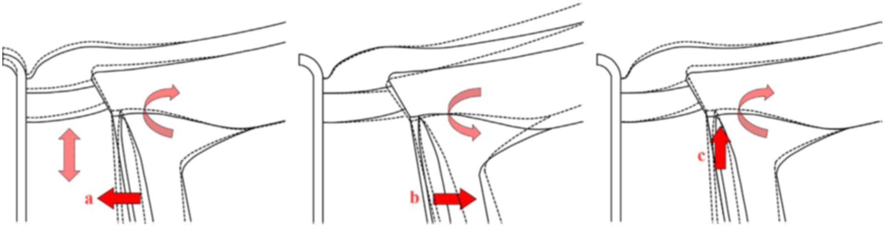
A schematic description of three mechanisms by which increases in CSFP cause ONH deformations. Undeformed ONH is shown with continuous lines, and deformed ONH with dashed lines. (a) CSFP acts inwardly compressing the pia mater and the retrolaminar neural tissue within. Due to the Poisson effect, lateral compression may cause expansion in the axial direction, increasing retrolaminar pressure (Morgan et al., 1995) “pushing” anteriorly on the lamina and causing clockwise rotation of the PPS. (b) CSFP acts outwardly on the dura mater away from the pia mater, causing the known distension of the dural sheath, (Killer et al., 2003) rotating the PPS counterclockwise, and displacing the periphery of the lamina posteriorly. (c) CSFP “pushes” the PPS anteriorly, causing flattening of the globe and clockwise rotation of the PPS, and displacing the periphery of the lamina anteriorly. Figure adapted from (Hua et al., 2018) with permission of the Association for Research in Vision and Ophthalmology.