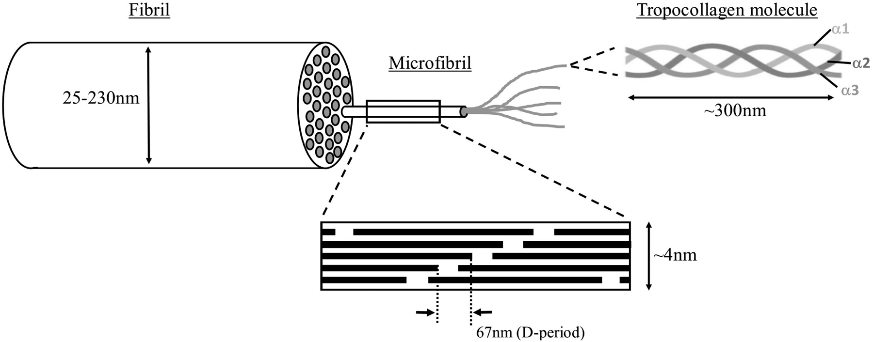
Figure 3:

The hierarchical structure of scleral collagen (not to scale). Five triple alpha-chain tropocollagen molecules assemble into microfibrils, in which the axial stagger of individual molecules leads to gap/overlap regions that define the 67nm axial D-period. Varying numbers of near-parallel microfibrils form collagen fibrils of diameters ranging from 25 to 230nm.The microfibrils are actually inclined by ~5° to the fibril axis, but this is not shown in this simplified diagram.
