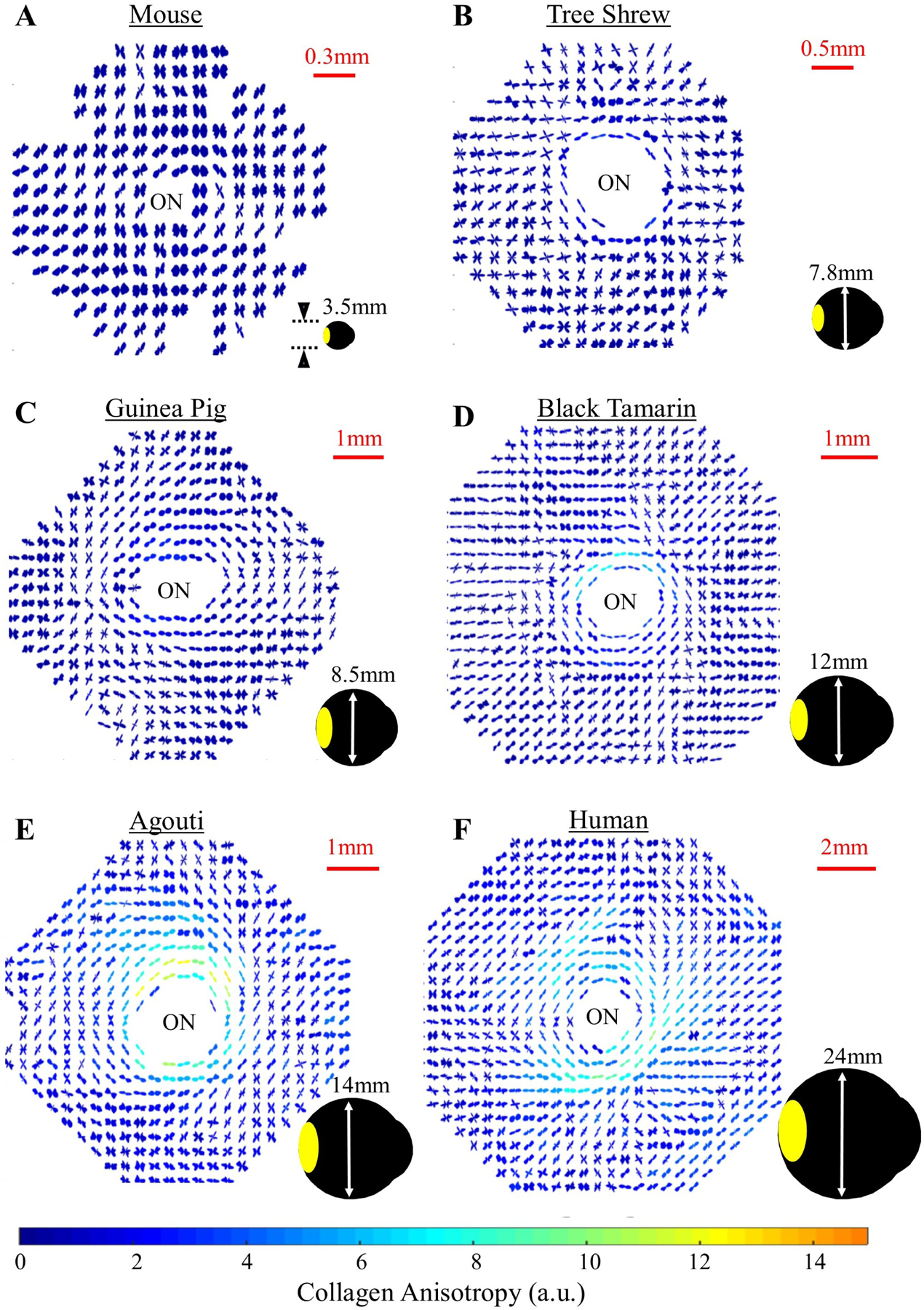
Figure 6:

Collagen microstructure of the posterior sclera across species. Polar vector plots of collagen fibril orientation in various mammal species (A–E) and humans (F), determined using WAXS. The shape of the individual plots indicates the preferred direction of collagen fibrils at that point in the tissue, while the plot colour scaling is indicative of the degree of anisotropy. Note that the circumferential collagen structure of the peripapillary sclera bordering the optic nerve (ON) is poorly defined in smaller mammals, but becomes gradually clearer with increasing eye size. The area covered by the WAXS maps is shown in yellow on the accompanying eye shadow diagrams.
