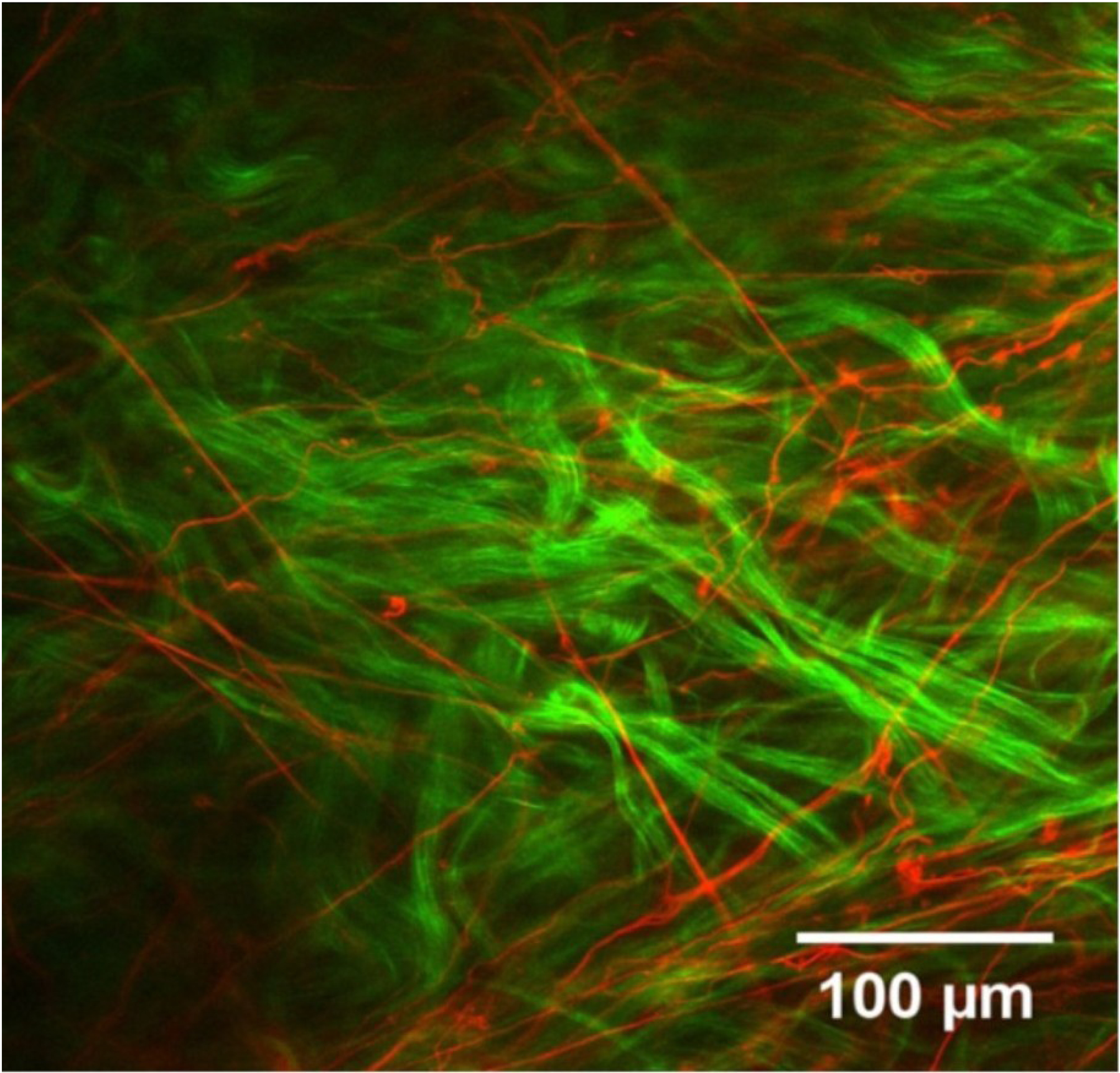
Figure 8:

Two-channel multiphoton microscopy image recorded from human episclera. The elastin fibre network (red) is revealed by TPF autofluorescence, and is shown alongside collagen fibril bundles (green) visualized concurrently with SHG imaging. Figure adapted from (Park et al., 2016) with permission of the Association for Research in Vision and Ophthalmology.
