FIG. 2.
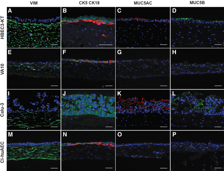
Immunofluorescent staining of vimentin, CK5, CK18, MUC5AC, and MUC5B in 3D airway mucosa tissue models. Vimentin is localized in the SIS of each cell line-based model (A, E, I, M). 3D tissue models based on HBEC3-KT, VA10, and Cl-huAEC show CK5 (red) and CK18 (green) expression in the epithelial compartment (B, F, N), whereas Calu-3-based models show only CK18 expression (J). MUC5AC and MUC5B are identified in HBEC3-KT- (C, D), VA10- (G, H), and Calu-3-based models (K, L), but are not visible in Cl-huAEC-based models (O, P). Scale bars: 50 μm. CK, cytokeratin; MUC, mucin; SIS, small intestinal submucosa.

