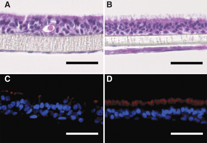
FIG. 5.
Morphological characterization of airway mucosa models on transwell inserts using primary airway fibroblasts and HBEC3-KT (A, C) or hAEC (MucilAir™; B, D). H&E stainings show differentiated epithelial cell layers (A, B) with beta tubulin-positive kinocilia (red fluorescence in C, D). Scale bars: 50 μm. hAEC, human primary airway epithelial cells.

