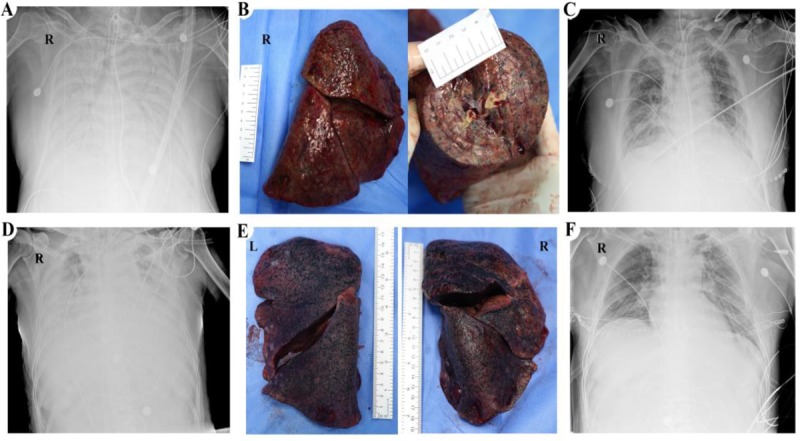
FIGURE 1.

Chest x-rays and gross photos of patients. (A) The first patient's chest x-ray on February 27. (B) Frontal view and cross-sectional appearance of the first patient's resected right lung. (C) The first patient's chest x-ray on four days after lung transplantation. (D) The second patient's chest x-ray on March 7. (E) Resected lungs of the second patient. (F) The second patient's chest x-ray on the next day of lung transplantation. L indicates left; R, right.
