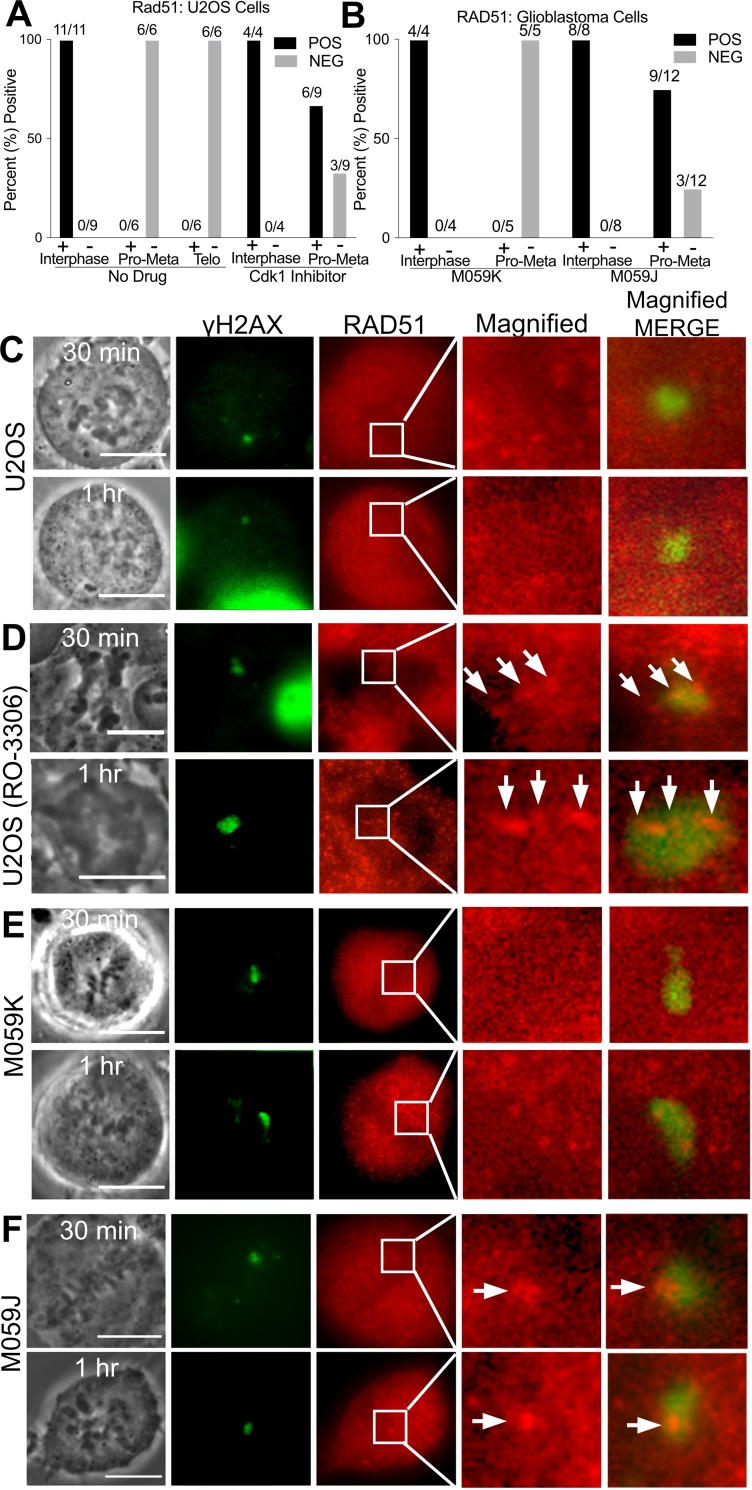
Fig 6. DNA damaged chromosome regions are devoid of RAD51.
(A) Percent of U2OS cells fixed 25–90 minutes post laser that are positive for RAD51 according to mitotic phase based on immunofluorescence staining data. CDK1 inhibition causes some cells to present mean pixel values above background, MPI = 53 ± 16 in six out of 9 cells. Corresponding box plot can be supplemental files. (B) DNA Pkcs deficient cells, (M059J) also show a greater likelihood of RAD51 above background levels when compared to U2OS. Nine out of twelve had positive RAD51, MPI = 121 ± 67. Corresponding box plot can be found in supplemental files. C) U2OS mitotic cells stained for γh2AX and RAD51. (D) U2OS treated with 10 μM CDK1 inhibitor underwent premature cytokinesis and chromosome de-condensation. Nevertheless, RAD51 co-localized with γh2AX and appears dotted along a track in an enlarged image of a boxed region shown on the RAD51 column. (E) The isogenic glioblastoma lines M059K and (F) M059J were DNA damaged with the laser and stained for γh2AX and RAD51. (F) M059J cells (deficient in DNA-PKcs) show RAD51 at the damage spot. Scale bar = 10μm.

