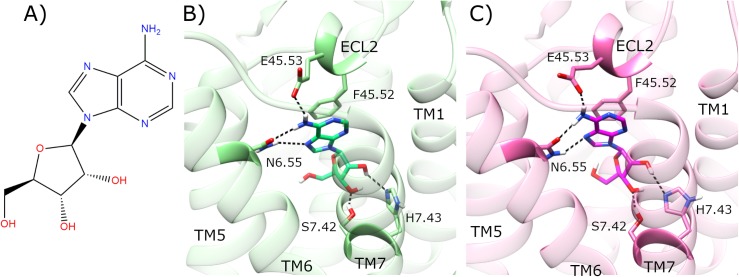
Fig 2. Docking of adenosine in the inactive crystal structure of adenosine A2a receptor (A2aR).
A) Molecular structure of adenosine. Comparison of B) co-crystallized adenosine (lime) in agonist-bound A2aR crystal structure (PDB entry: 2YDO, light green), and C) docked adenosine (magenta) in the inactive crystal structure of A2aR (PDB entry: 4EIY, pink). Selected residues participating in ligand binding are displayed. ECL2 and TM helices 1, 5–7 are labelled.

