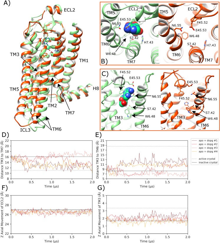
Fig 5. Transition towards an alternative intermediate conformation of apo A2aR in a DOPG membrane.
A) Comparison of the intermediate state crystal structure of A2aR (PDB entry: 2YDO, light green) with bound adenosine and an MD-generated apo conformation achieved within a DOPG membrane (orange, belonging to replica #1 from 1.6 μs), showing B) and C) selected residues delineating the orthosteric pocket. ECL2 and TM helices are labelled where applicable. D) Distance between TM3-TM7 (from Cα atoms of R1023.50 and Y2887.53, respectively) during MD simulations, starting from the inactive crystal structure (PDB entry: 4EIY). E) Distance between TM3-TM6 (from Cα atoms of R1023.50 and E2286.30, respectively). F) and G) Vertical movement of ECL2 and TM3, respectively. MD simulations are performed in quadruplicate. Corresponding flat-lines show the observed distance in the active (PDB entry: 6GDG) and inactive (PDB entry: 4EIY) A2aR crystal structures.

