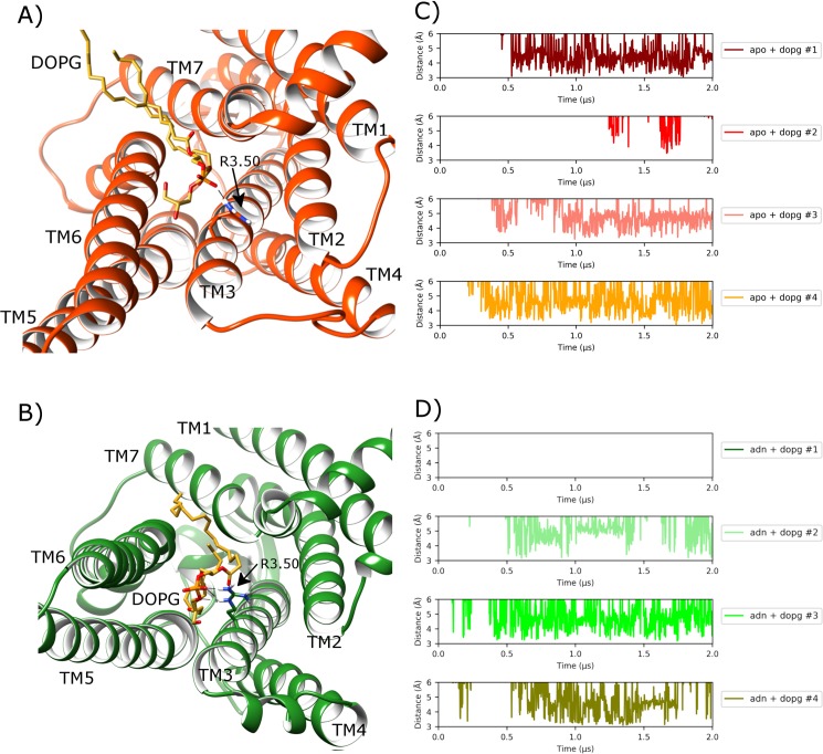
Fig 7. Protein-lipid allosteric interaction with the ionic-lock in MD simulations of A2aR in a DOPG membrane.
A) Intracellular view of A2aR where ionic-lock residue R1023.50 electrostatically interacts with a DOPG lipid, which intrudes between TM6 and TM7 into the G protein binding-site in apo state (orange) (belonging to replica #4 from 1.8 μs), and B) with bound adenosine (green) (belonging to replica #2 from 1.6 μs). Protein-lipid interaction distance over time between R1023.50 sidechain and lipid phosphate group in four replicas of A2aR in DOPG membrane in C) apo state and D) adenosine-bound (ADN), respectively.

