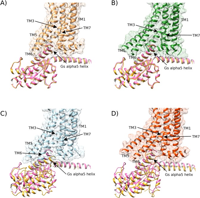
Fig 11. The MD-generated receptor conformation of A2aR bound to adenosine in DOPG membrane is able to bind co-crystallized Gs-alpha protein in the same way as the active-state crystal structure (PDB id: 6GDG).
A) The crystal structure of the active state of A2aR (brown) bound to its co-crystallized Gs-alpha protein (pink, PDB id: 6GDG) and its re-docked Gs-alpha subunit superimposed (gold). B) MD-generated active-like conformation of A2aR bound to adenosine in a DOPG membrane (green, belonging to replica #2 from 1.6 μs) docks Gs-alpha protein (gold) in similar fashion to the active crystal (pink). C) Intermediate conformation of A2aR, bound to adenosine in DOPC membrane (blue, belonging to replica #4 from 1.3 μs) fails to properly dock Gs-alpha protein (gold) compared to its active crystal position (pink). D) Intermediate conformation of apo A2aR in DOPG membrane (red, belonging to replica #1 from 1.6 μs) partially docks Gs-alpha protein (gold) compared to its active crystal position (pink).

