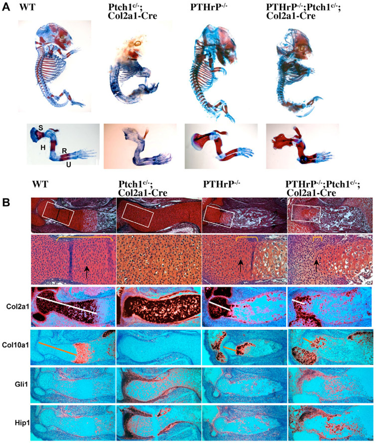
Fig. 2. Cell autonomous upregulation of Hh signaling in the absence of PTHrP accelerates chondrocyte hypertrophy.
(A) Skeletal preparation of embryos at E14.5. Alizarin Red stains mineralized cartilage and bone tissues; Alcian Blue stains unmineralized cartilage. A higher magnification view of the forelimb is shown in the lower panel. S, scapula; H, humerus; R, radius; U, ulna. (B) Serial sections of distal humerus were stained with Safranin O and hybridized with indicated 35S labeled riboprobes. The boxed articular and columnar chondrocytes regions are shown at higher magnification in the panels below. Columnar chondrocytes are indicated by yellow brackets and arrows. Both Ptch1c/−;Col2a1-Cre and PTHrP−/−;Ptch1c/−;Col2a1-Cre mutants show strong upregulation of Gli1 and Hip1, downstream target genes of Hh signaling, in the perichondrium and synovial joint. The Col2a1-expressing region (white line) is reduced and the Col10a1-expressing domain (yellow line) is closer to the joint in the PTHrP−/−;Ptch1c/−;Col2a1-Cre mutant.

