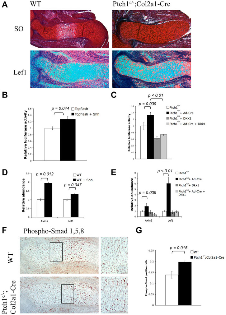
Fig. 5. Hedgehog signaling activates downstream targets of canonical Wnt signaling both in vivo and in vitro.
(A) Serial sections of E14.5 distal humerus were stained by Safranin O and hybridized with a 35S labeled Lef1 probe. Lef1 expression was strongly upregulated in the cartilage of Ptch1c/−;Col2a1-Cre mutants. (B,C) Dual luciferase assay of primary chondrocytes isolated from wild-type newborn pups. Primary chondrocytes were nucleofected with Topflash reporter vectors as a read out for canonical Wnt signaling. Recombinant Shh or Dkk1 protein was added after serum starvation and luciferase activity was measured 24 hours later. Shh treatment or Cre-adenovirus infection of the Ptch1c/c primary chondrocytes activated TOPFLASH activity. Such activation was diminished by Dkk1. (D,E) Quantitative RT-PCR was performed using RNA isolated from primary chondrocytes. Both Axin2 and Lef1 were significantly increased in Shh-treated primary chondrocytes or Cre-adenovirus-infected Ptch1c/c primary chondrocytes compared with untreated samples. Dkk1 treatment blocked the effect of Hh signaling. (F) Immunohistochemistry of E15.5 limb sections (distal ulna) with antibodies against phospho-Smad1, 5 and 8. More phospho-Smad-positive cells were found in the cartilage of the Ptch1c/−;Col2a1-Cre mutant embryos. Boxed region of columnar/prehypertrophic chondrocytes is shown at a higher magnification on the right-hand side. (G) Statistical analysis of phospho-Smad-positive cells in the boxed region of the cartilage. Three samples in the boxed area were counted, and the mean±s.d. are shown. Student′s t-test, P<0.05.

