FIGURE 1 -.

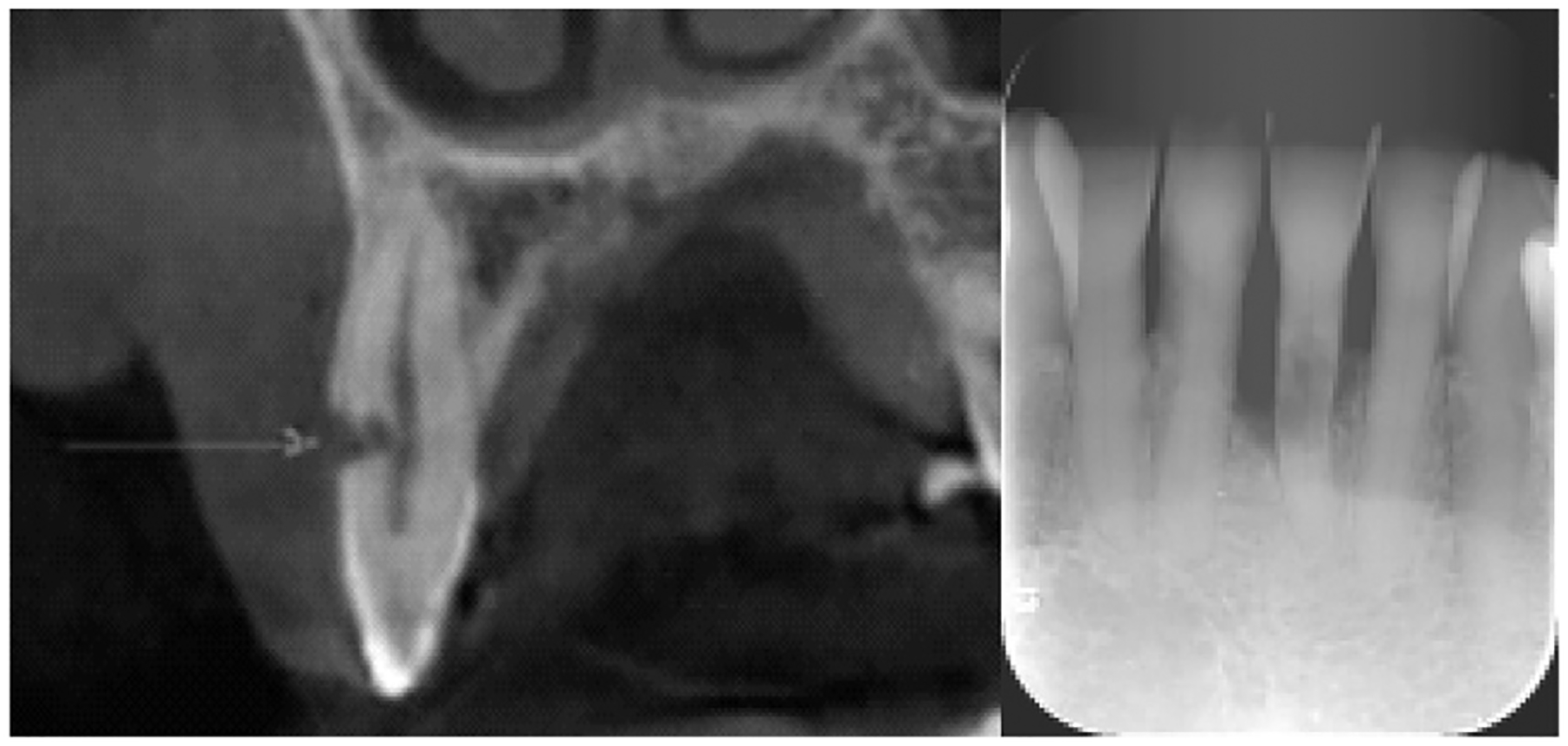
Representative ECR lesions shown on radiography. ECR is present on (left) tooth #6 and (right) tooth #24. The arrow indicates an ECR lesion at the buccal side of #6 on radiography.

Representative ECR lesions shown on radiography. ECR is present on (left) tooth #6 and (right) tooth #24. The arrow indicates an ECR lesion at the buccal side of #6 on radiography.