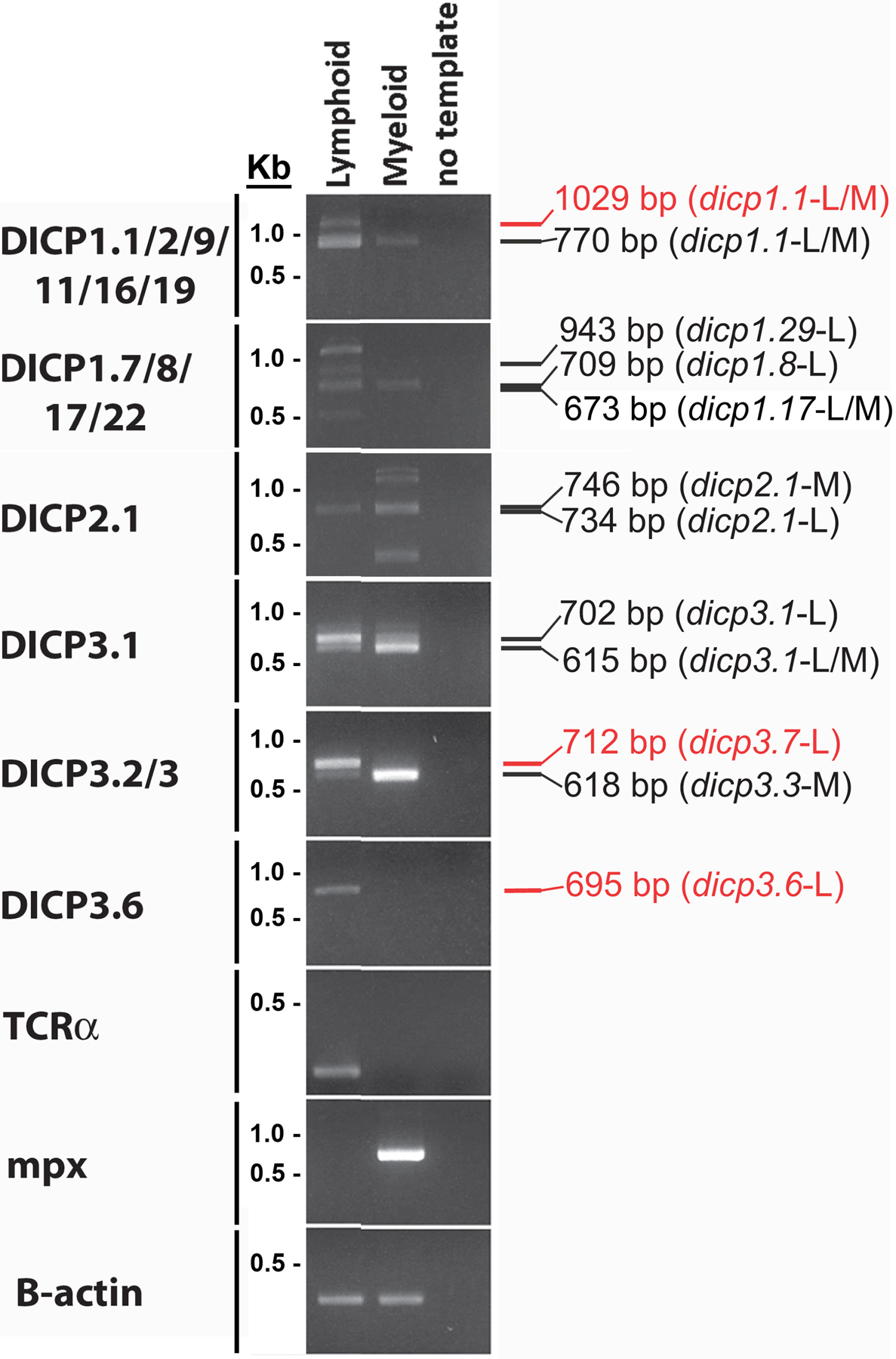
Fig. 3. DICP gene expression in myeloid and lymphoid cells.

Kidney cells from five adult EKW zebrafish were pooled and lymphoid and myeloid cells sorted by flow cytometry. DICP transcripts were amplified by RT-PCR with Titanium Taq DNA polymerase and all amplicons detected were cloned and sequenced to confirm their identity. The size and identity of recovered amplicons is listed on the right of the gel image; red text indicates nonfunctional transcripts. RT-PCR of myeloperoxidase (mpx) provides a positive control for myeloid cells and TCR-α provides a positive control for T lymphocytes. β-actin expression was used as a reference for cDNA quantity and quality.
