Fig. 7.
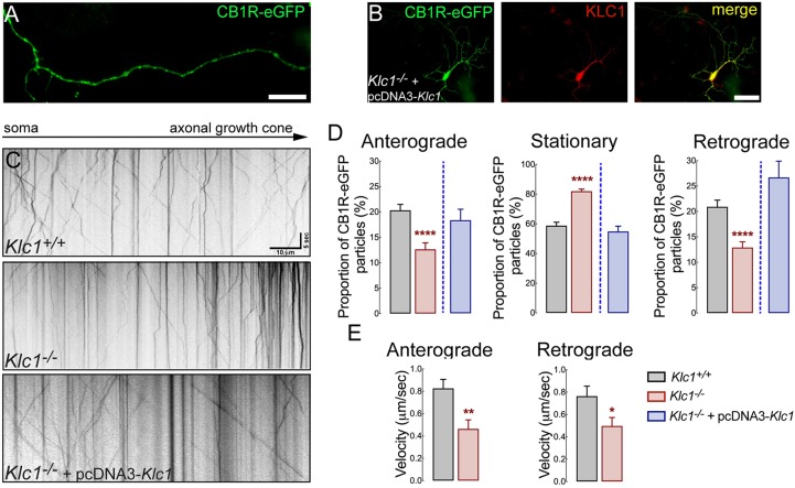
CB1R axonal transport is impaired in Klc1−/− neurons. (A) Epifluorescence image obtained from a live-cell movie of 7-8 DIV hippocampal neurons expressing CB1R-eGFP. (B) Hippocampal neurons co-transfected with CB1R-eGFP+pcDNA-KLC1 and immunostained for KLC1 showing KLC1 overexpression. (C) Kymographs of CB1R-eGFP transfected neurons obtained from live-cell imaging of Klc1+/+, Klc1−/−, and Klc1−/−+pcDNA-KLC1 hippocampal cultures. (D,E) Quantification of anterograde (Anter), stationary (Stat) and retrograde (Retro) proportion of CB1R-eGFP vesicles (D), and average velocities for anterograde and retrograde CB1R-eGFP vesicles (E). Data are mean±s.e.m. Klc1+/+, n=52; Klc1−/−, n=43; Klc1−/−+pcDNA-KLC1, n=34 neurons from three independent experiments. *P<0.01, **P<0.001, ****P<0.0001; Student's t-test. Scale bars: 20 μm (A); 50 μm (B).

