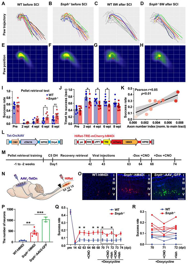
Figure 3. Regenerated CST Axons Contribute to Recovery in Manual Dexterity.

(A-D) Paw motion trajectories in the reaching phase of pellet retrieval test in WT and Snph−/− mice before and 8 weeks after the C5 DH (8 wpi). Successful reaches are colored and unsuccessful attempts are grey.
(E-H) Heat maps of paw spatial positions in the grasping phase relative to the food pellet (red spot) in WT and Snph−/− mice before and at 8 wpi.
(I-K) Analysis of the success rate of skilled reaching (I) and time to successful retrieval (J) shows that Snph−/− mice outperformed WT mice in the dexterity task. (K) Correlation between success rate and BDA-labeled CST axon number index.
(L-N) Schematics showing dual viral vectors (L) and experimental design (M) of DREADDs-mediated silencing of CST neurons (N).
(O) Labeling of corticospinal neurons in the motor cortex that received dual viral or AAV2-GFP infection.
(P) Total number of dual-virus infected corticospinal neurons in WT and Snph−/− mice, as well as corticospinal neurons infected by AAV2-GFP virus in Snph−/− mice.
(Q, R) Changes of forelimb dexterity in pellet retrieval test via administration of CNO either before or after induction of hM4Di expression with doxycycline treatment.
Data were presented as mean ± sem. n = 10 mice/group in pellet retrieval test (A-K); n = 5-6 mice/group in DREADDs study (N-R). Two-way ANOVA with Bonferroni post hoc test. * P < 0.05, ** P < 0.01, *** P < 0.001. Scale bar: 200 μm (O). (Also see Figure S2).
