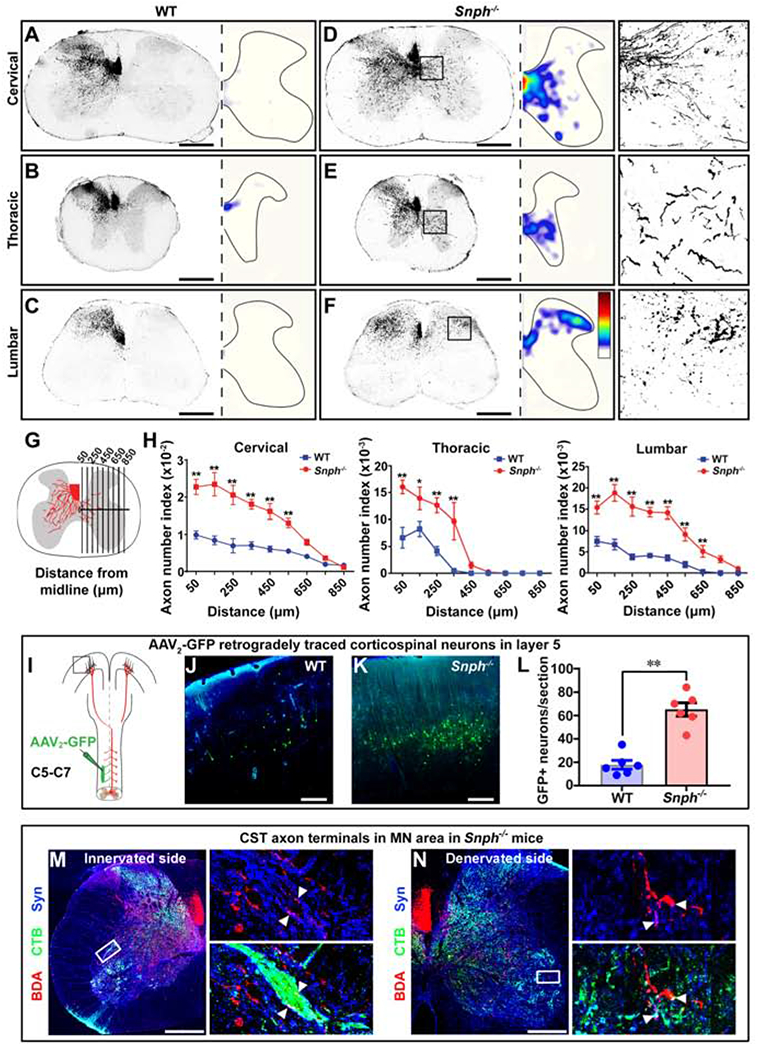
Figure 4. Snph−/− Mice Display Enhanced Compensatory CST Spouting after Unilateral Pyramidotomy.

(A-F) Cross-sectional images showing midline-crossing of BDA-labeled CST axons at cervical, thoracic, and lumbar levels following unilateral (left) pyramidotomy. The distribution of axonal sprouting into the denervated side was converted to heatmaps; red represents the highest numbers of axon pixels, blue represents the lowest, and white represents background (D-F). Right panels in D-F: details of boxed areas showing BDA-labeled CST terminal sprouting at denervated regions of the cervical, thoracic, and lumbar cords, respectively, in a Snph−/− mouse.
(G, H) Schematic (G) and analysis (H) showing BDA-labeled midline-crossing CST axons.
(I) Unilateral injection of AAV2-GFP into the denervated (right) side of the C5-C7 spinal intermediate gray matter to retrogradely label corresponding corticospinal neurons in the motor cortex.
(J-L) Images of boxed area in I (J, K) and analysis (L) showing GFP-labeled CST neurons in layer V of WT and Snph−/− mice.
(M, N) Close apposition of BDA-labeled CST (red), synaptophysin (Syn)-labeled presynaptic terminals (blue), and CTB-labeled MNs (green) in both innervated (M) and denervated (N) spinal cord in Snph−/− mice. Left panels: high magnifications of boxed areas in M and N, respectively. Arrowheads indicate triple-positive appositions.
Data were presented as mean ± sem. n = 6 mice/group (H, L). Unpaired two-tailed Student’s t-test. * P < 0.05, ** P < 0.01. Scale bars: 500 μm (A-F), 250 μm (J, K), 400 μm (M, N). (Also see Figure S3)
